Do you require any assistance? Simply reserve your appointment online below
Radiology Services in Kenya
Patient – Centered Care
Nairobi Spine Orthopaedic Center in Kenya is a recognized leader in the delivery of medical imaging services. We offer a comprehensive range of radiology services using the latest technology to provide our patients and their referring physicians with superior diagnostic evaluation in a comfortable and welcoming environment.
Our imaging and radiology experts use advanced technologies to help diagnose and treat various conditions.
Comprehensive Radiology Procedures
The diagnostic equipment at Nairobi Spine Orthopedic Center features the latest in advanced, high-resolution digital imaging technology enabling our radiologists to diagnose and treat a variety of injuries, conditions and diseases. .
No matter the area of the body being examined, our physicians can quickly and easily have access to the images needed for the diagnosis and treatment of our patients.
Our comprehensive radiological procedures include open MRI,CT scans,fluoroscopy, ultrasound, bone density screenings, and other interventional radiology procedures.
Meet Your Radiologists
ulti-modality center for all your imaging needs. Our staff and physicians get you in and out of the center with minimal delay, and make your study available to your referring physician almost as soon as your feet hit the ground. Nairobi Spine & Orthopedic Center is the leader in orthopedic care that you can trust.
Radiology Services at NSOC
The following radiology services are available at NSOC, designed to better meet your needs.
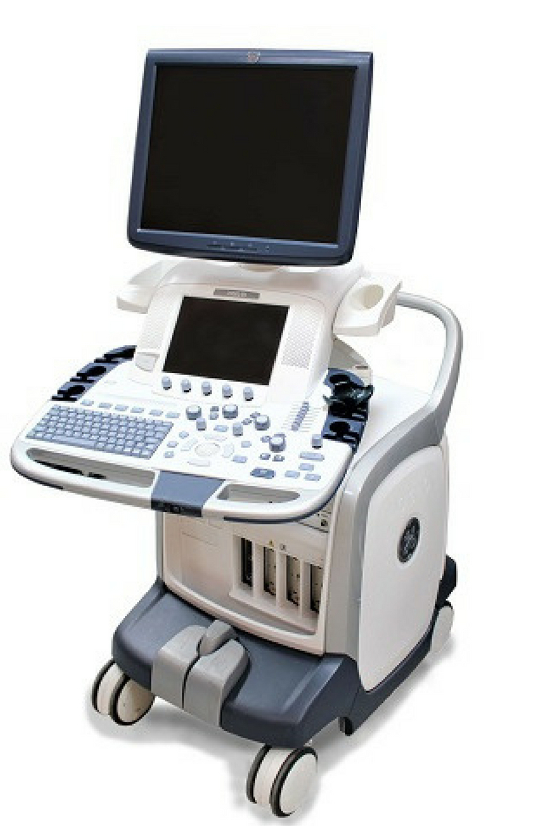
Diagnostic Ultrasound
Dedicated NSOC physicians in-office ultrasound provides images of muscles, tendons, ligaments, joints, and soft tissue throughout the body. It can be used to diagnose and treat a number of musculoskeletal conditions.
Ultrasound can be used to diagnose tendon tears, such as shoulder rotator cuff tears or Achilles tendon tears, abnormalities of the muscle, bleeding or other fluid collections within the muscles and joints, joint effusion or synovial thickening.
Ultrasounds are advantageous because, they are non-invasive, usually pain-free, no ionizing radiation, offer excellent imaging of soft tissues, provides real-time imaging, not affected by cardiac pacemakers or implants and the patient is able to resume normal activities immediately following an ultrasound examination
Fluoroscopy
Fluoroscopy is a form of diagnostic radiology that enables our physicians, with or without the aid of a contrast agent, to visualize area of concern via the x-ray.
This contrast agent allows the structures in the body to be viewed clearly, in real-time, or “live” on a television monitor or screen. Fluoroscopy, also called C-Arm, ensures optimal application flexibility and customizable x-ray functionality.

Computed Tomography
Computed tomography (CT) is an imaging tool that combines x-rays with computer technology to produce a more detailed, cross-sectional image of your body. A CT scan lets your doctor see the size, shape, and position of structures that are deep inside your body, such as organs, tissues, or tumors. Tell your doctor if you are pregnant before undergoing a CT scan.
While on a CT scan,You lie as motionless as possible on a table that slides into the center of the cylinder-like CT scanner. The process is painless. An x-ray tube slowly rotates around you, taking many pictures from all directions.
A computer combines the images to produce a clear, two-dimensional view on a television screen.
Magnetic resonance imaging
Magnetic resonance imaging (MRI) is another modern diagnostic imaging technique that produces cross-sectional images of your body. Unlike CT scans, MRI works without radiation. The MRI tool uses magnetic fields and a sophisticated computer to take high-resolution pictures of your bones and soft tissues.
You lie as motionless as possible on a table that slides into the tube-shaped MRI scanner. The MRI creates a magnetic field around you and then pulses radio waves to the area of your body to be pictured. The radio waves cause your tissues to resonate.
A computer records the rate at which your body’s various parts (tendons, ligaments, nerves) give off these vibrations, and translates the data into a detailed, two-dimensional picture. You will not feel any pain while undergoing an MRI, but the machine may be noisy.
An MRI may help your doctor to diagnose your torn knee ligaments and cartilage, torn rotator cuffs, and other problems.


Bone Densitometry (DEXA)
This allows for scan analysis of the spine, hip and forearm determining the bone density of our patients.
DEXA allows the healthcare provider to visualize unknown fractures in the spine using an Instant Vertebral Analysis (IVA). It is a painless exam that instantly produces a report for diagnostic interpretation.
The provider can see the patient as soon as the exam is completed. A DEXA scan is a safe non-invasive exam that is done in the comfort of our office.
It uses a low dose of radiation to produce accurate results. This simple test can detect the beginning of bone loss as well as advanced cases of osteoporosis.
DEXA scan is the first step toward healthier bones for the future.

